Table of Contents
(tap to open/close)
Human Reproduction
- Humans reproduce sexually and give birth to live young ones, therefore they are viviparous.
- Human reproduction involves a series of well-defined events that occur after puberty.
Key Reproductive Events
- Gametogenesis: Formation of sperms in males and ovum in females.
- Insemination: Transfer of sperms into the female genital tract.
- Fertilisation: Fusion of male and female gametes to form a zygote.
- Blastocyst Formation: Development of the zygote into a blastocyst.
- Implantation: Attachment of the blastocyst to the uterine wall.
- Gestation: Development of the embryo inside the uterus.
- Parturition: Delivery of the baby.
1.1 The Male Reproductive System

showing reproductive system
- Location:
- The male reproductive system is located in the pelvic region.
- Major Components:
- Primary Sex Organ: A pair of testes.
- Secondary Structures: Accessory ducts, accessory glands, and external genitalia (penis and scrotum).

(part of testis is open to show inner details)
Testes
- Position and Temperature Regulation:
- The testes are located outside the abdominal cavity within the scrotum.
- This position maintains the testes at a temperature about 2–2.5°C lower than body temperature, which is essential for sperm production.
Functions of Testes
- Production of sperms.
- Secretion of male sex hormones.
Structure of Testes
- Each testis is oval in shape, about 4–5 cm long and 2–3 cm wide.
- It is covered by a dense protective covering.
- Each testis contains about 250 compartments called testicular lobules.
- Each lobule has 1–3 highly coiled seminiferous tubules where sperms are produced.
Seminiferous Tubules
- Lining Cells:
- Male Germ Cells (Spermatogonia): Undergo meiosis to form sperms.
- Sertoli Cells: Provide nutrition and support to developing sperms, hence called nurse cells.
- Interstitial Spaces:
- Present between seminiferous tubules.
- Contain blood vessels and Leydig cells.
- Leydig Cells: Secrete testicular hormones called androgens (mainly testosterone).


Male Accessory Ducts
- Types:
- Rete testis
- Vasa efferentia
- Epididymis
- Vas deferens
- Functions:
- Transport sperms from testes to the urethra.
- Store sperms temporarily and provide nourishment.
Epididymis
- Differentiated into caput, corpus, and cauda epididymis.
- Stores sperms and allows their maturation.
Vas Deferens
- Carries sperms from epididymis.
- Joins the duct of seminal vesicle to form the ejaculatory duct.
Ejaculatory Duct
- Carries sperms and secretions of seminal vesicles into the urethra.
External Genitalia
Penis
- Acts as the male copulatory organ.
- Also serves as a passage for urine and semen.
- Made of erectile spongy tissue that becomes erect when filled with blood.
- Glans Penis
- Enlarged terminal part of the penis.
- Covered by a loose fold of skin called the foreskin or prepuce.
Male Accessory Glands
- Types
- Seminal vesicles
- Prostate gland
- Bulbourethral (Cowper’s) glands
- Functions
- Their secretions together form seminal plasma.
- Provide nutrition, lubrication, and protection to sperms.
- Seminal Vesicles
- Produce an alkaline secretion rich in fructose, calcium, enzymes, and prostaglandins.
- Fructose provides energy to sperms.
- Prostaglandins help in sperm movement inside the female reproductive tract.
- Prostate Gland
- Secretes a slightly alkaline milky fluid.
- Activates and nourishes sperms.
- Helps neutralise acidity of the vaginal tract.
- Bulbourethral Glands
- Secrete alkaline mucus.
- Neutralise acidity of urine in urethra.
- Lubricate the penis during copulation.
Semen
- Definition:
- Semen is a mixture of sperms and secretions of seminal vesicles, prostate gland, and bulbourethral glands.
- Characteristics:
- Ejected during ejaculation.
- One ejaculation may contain 200–300 million sperms.
- Alkaline in nature (pH about 7.35–7.50).
- Provides a fluid medium for sperm transport and survival.
Related Disorders (Important)
- Cryptorchidism
- Failure of one or both testes to descend into the scrotum.
- Caused by deficient testosterone secretion during fetal life.
- Inguinal Hernia
- Protrusion of a part of intestine into the scrotum due to tearing of inguinal tissue.
1.2 The Female Reproductive System
The female reproductive system is structurally and functionally designed to support ovulation, fertilisation, pregnancy, childbirth, and child care.

reproductive system
Main Components
- The female reproductive system consists of:
- Ovaries
- Oviducts (Fallopian tubes)
- Uterus
- Cervix
- Vagina
- External genitalia
- Mammary glands
All these structures are located in the pelvic region and work in coordination.
Ovaries
- Nature and Position:
- Primary female sex organs.
- Present on either side of the lower abdomen in the pelvic cavity.
- Each ovary is about 2–4 cm long.
- Functions
- Produce female gametes (ova).
- Secrete ovarian hormones such as oestrogen and progesterone.
- Structure of Ovary
- Covered externally by a thin germinal epithelium.
- Below it lies the tunica albuginea.
- Interior region is called ovarian stroma, which is differentiated into:
- Cortex: Outer dense region containing developing follicles.
- Medulla: Inner region with blood vessels, nerves, elastic fibres and smooth muscles.
- Attachments
- Ovary is attached to the uterus by the ovarian ligament.
- Also connected to the pelvic wall by suspensory ligaments.
Oviducts (Fallopian Tubes)
- General Features
- Paired muscular tubes, each about 10–12 cm long.
- Extend from the ovaries to the uterus.
- Lined internally by ciliated epithelium.
- Movement of ovum occurs by ciliary action and peristalsis.
Parts of Oviduct
- Infundibulum:
- Funnel-shaped structure near the ovary.
- Possesses finger-like projections called fimbriae which collect the ovum after ovulation.
- Ampulla:
- Wider middle portion of the oviduct.
- Isthmus:
- Narrow terminal part that joins the uterus.
Site of Fertilisation
- Fertilisation occurs at the ampullary–isthmic junction of the fallopian tube.
Uterus (Womb)
- General Features:
- Single, hollow, muscular organ.
- Inverted pear-shaped.
- Supported by ligaments attached to the pelvic wall.
- Functions:
- Site of implantation of blastocyst.
- Supports development of embryo and foetus.
- Participates in childbirth through muscular contractions.
Parts and Layers of Uterine Wall
- Perimetrium:
- Outer thin membranous layer.
- Myometrium:
- Middle thick layer of smooth muscles.
- Shows strong contractions during parturition.
- Endometrium:
- Inner glandular layer lining the uterine cavity.
- Undergoes cyclical changes during the menstrual cycle.
Cervix
- Description:
- Lower narrow part of the uterus.
- Connects uterus to the vagina.
- Cervical Canal:
- Cavity of the cervix is called the cervical canal.
- Cervical canal together with vagina forms the birth canal.
Vagina
- Structure:
- Muscular tube about 10 cm long.
- Extends from cervix to outside of the body.
- Functions:
- Receives penis during copulation.
- Provides passage for menstrual flow.
- Acts as birth canal during childbirth.
Hymen
- Thin membrane that partially covers the vaginal opening.
- May rupture due to physical activities like cycling or sports.
- Its presence or absence is not a reliable indicator of virginity.

External Genitalia
Components
- Mons Pubis:
- Fatty cushion covered with skin and pubic hair
- Labia Majora:
- Large fleshy folds extending from mons pubis and surrounding vaginal opening.
- Labia Minora:
- Thin paired folds present under labia majora.
- Clitoris:
- Small, cap/finger-like, highly sensitive structure.
- Located at the upper junction of labia minora above the urethral opening.
Mammary Glands

- Nature and Function:
- Paired structures called breasts.
- Function is milk production (lactation) to nourish newborn.
- Structure of Mammary Glands:
- Each breast contains glandular tissue and variable amount of fat.
- Glandular tissue is divided into 15–20 mammary lobes.
- Alveoli:
- Each lobe contains clusters of secretory cells called alveoli.
- Alveoli secrete milk and store it in their lumens.
- Milk Pathway
- Alveoli → Mammary tubules → Mammary ducts → Mammary ampulla → Lactiferous duct → Outside
1.3 Gametogenesis
- Definition:
- Gametogenesis is the process of formation of male and female gametes in humans.
Primary Sex Organs Involved
- Testis: Produce male gametes (sperms).
- Ovaries: Produce female gametes (ova).
Spermatogenesis (Formation of Sperms)

seminiferous tubule (enlarged)
- Definition:
- Spermatogenesis is the process of formation of male gametes (sperms) from spermatogonia.
- Site:
- Occurs in the seminiferous tubules of the testes.
- Time of Initiation:
- Begins at puberty and continues throughout reproductive life.
Phases of Spermatogenesis
- Multiplication Phase
- Undifferentiated male germ cells called spermatogonia (46 chromosome) divide repeatedly by mitosis.
- Some spermatogonia remain as stem cells to maintain continuity of spermatogenesis.
- Growth Phase
- Some spermatogonia grow in size and become primary spermatocytes.
- Primary spermatocytes are diploid (46 chromosomes).
- Maturation Phase
- Primary spermatocyte undergoes first meiotic (reduction) division to form two secondary spermatocytes.
- Secondary spermatocytes are haploid (23 chromosomes).
- Each secondary spermatocyte undergoes second meiotic (equational) division to form two spermatids.
- Thus, one primary spermatocyte produces four haploid spermatids (23 ch.).
Note – The formation of primary spermatocytes from spermatogonia is called spermatocytogenesis.
Spermiogenesis
- Definition:
- Spermiogenesis is the transformation of spermatids into mature spermatozoa (sperms).
- Outcome:
- Spermatids undergo structural differentiation to form head, neck, middle piece and tail.
Spermiation
- Definition:
- After spermiogenesis, the heads of spermatozoa remain embedded in Sertoli cells.
- They are finally released into the lumen of seminiferous tubules.
- This release of sperms from Sertoli cells is called spermiation.
Structure of a Sperm
General Features
- Sperm is microscopic and motile.
- Entire body is covered by a plasma membrane.
Parts of a Sperm
- Head
- Contains an elongated haploid nucleus.
- Anterior part is covered by acrosome.
- Acrosome contains hydrolytic enzymes such as acrosin and hyaluronidase.
- These enzymes help in penetration of ovum during fertilisation.
- Neck
- Short connecting region between head and middle piece.
- Middle Piece
- Contains numerous mitochondria arranged spirally.
- Provides energy for movement of tail.
- Tail
- Long and slender structure.
- Responsible for motility of sperm.

Semen
- Definition
- Semen is a mixture of sperms and seminal plasma.
- Components
- Sperms produced in testes.
- Seminal plasma secreted by seminal vesicles, prostate gland and bulbourethral glands.
- Functions
- Provides nourishment to sperms.
- Facilitates transport and motility of sperms.
- Neutralises acidity of urethra and vaginal tract.
Normal Male Fertility
- A normal male requires:
- About 60% structurally normal sperms.
- About 40% motile sperms in semen.
Hormonal control of Spermatogenesis
- Role of Hypothalamus:
- At puberty, hypothalamus releases Gonadotropin Releasing Hormone (GnRH).
- Role of Anterior Pituitary: GnRH stimulates anterior pituitary to secrete:
- Luteinizing Hormone (LH).
- Follicle Stimulating Hormone (FSH).
- Role of LH:
- LH acts on Leydig cells.
- Leydig cells secrete androgens (mainly testosterone).
- Role of FSH:
- FSH acts on Sertoli cells.
- Stimulates spermiogenesis.
- Sertoli cells also secrete:
- Androgen Binding Protein (ABP) – concentrates testosterone in seminiferous tubules.
- Inhibin – inhibits FSH secretion.
Feedback Regulation
- Testosterone inhibits LH secretion.
- Inhibin inhibits FSH secretion.
- This maintains hormonal balance.
Oogenesis (Formation of Ova)
- Definition
- Oogenesis is the process of formation of a mature female gamete (ovum) from oogonia.
- Location
- Occurs in the ovaries.
- Time of Initiation
- Oogenesis begins during embryonic development of the female fetus.
- No new oogonia are formed after birth.
Stages of Oogenesis
- Formation of Oogonia
- During fetal life, millions of oogonia (diploid, 46 chromosomes) are formed in each ovary.
- Oogonia multiply by mitosis.
- Formation of Primary Oocytes
- Oogonia differentiate into primary oocytes (diploid, 46 chromosomes).
- Primary oocytes enter meiosis I but get arrested at prophase I (diplotene stage).
- This arrested stage may last for years until puberty.
Each primary oocyte gets surrounded by a single layer of granulosa cells and is called a primary follicle.
Follicular Development After Puberty
- Primary Follicle:
- Primary oocyte surrounded by a single layer of granulosa cells.
- Secondary Follicle:
- Primary follicle surrounded by multiple layers of granulosa cells.
- Theca layer differentiates around granulosa cells.
- Tertiary Follicle:
- Characterised by the presence of a fluid-filled cavity called antrum.
- Primary oocyte increases in size.
Completion of First Meiotic Division
- At the tertiary follicle stage, the primary oocyte completes meiosis I.
- This division is unequal and produces:
- One large haploid secondary oocyte (23 chromosomes).
- One small first polar body.
Graafian Follicle and Ovulation
- The tertiary follicle matures into a Graafian follicle.
- Graafian follicle releases the secondary oocyte during ovulation.
In humans, the ovum is released in the secondary oocyte stage.
Completion of Meiosis II
- The secondary oocyte enters meiosis II but gets arrested at metaphase II.
- Meiosis II is completed only after sperm entry (fertilisation) in the fallopian tube.
Final Outcome
- From one oogonium, the following are formed:
- One functional ovum.
- Three polar bodies (non-functional).
Important Points
- Majority of primary follicles degenerate from birth to puberty.
- At puberty, only about 60,000–80,000 primary follicles remain in each ovary.
- Ovum is the largest cell in the human body.
- Ovum retains maximum cytoplasm to support early embryonic development.

Hormonal Control of Oogenesis
- Hypothalamus:
- Secretes Gonadotropin Releasing Hormone (GnRH).
- Anterior Pituitary: GnRH stimulates secretion of:
- Follicle Stimulating Hormone (FSH).
- Luteinizing Hormone (LH).
- Role of FSH:
- Stimulates growth of ovarian follicles.
- Promotes oocyte development and completion of meiosis I.
- Stimulates estrogen secretion.
- Role of LH:
- Induces ovulation.
- Stimulates corpus luteum to secrete progesterone.
Feedback Regulation
- Rising progesterone inhibits GnRH secretion.
- This reduces LH and FSH release.
Vitellogenesis
- Vitellogenesis is the process of yolk deposition in the primary oocyte.
- Occurs during prophase I of meiosis.
Key Differences Between Spermatogenesis and Oogenesis
| Feature | Spermatogenesis | Oogenesis |
|---|---|---|
| Process Initiation | Begins at puberty | Begins during fetal life but pauses temporarily (arrest). |
| Process Continuity | Continuous process | resumes at puberty |
| Site | Testes | Ovaries |
| End Products | Four spermatozoa (from one spermatogonium) | One ovum + polar bodies (from one oogonium) |
| Duration | About 64 days | Spans several years |
| Cytokinesis | Equal cytokinesis | Unequal cytokinesis |
| Gamete Size | Small with minimal cytoplasm | Large with abundant cytoplasm |
| Meiotic Division | Symmetrical | Asymmetrical (producing one large ovum and smaller polar bodies) |
| Number of Gametes | Millions produced continuously | One ovum released per menstrual cycle |
| Hormonal Control | Testosterone, FSH, LH | Estrogen, progesterone, FSH, LH |

1.4 Menstrual Cycle
- Definition:
- The menstrual cycle is the reproductive cycle in female primates, including humans.
- Menarche:
- Menarche is the first menstruation that occurs at puberty.
- Cycle Length:
- The average length of the menstrual cycle is about 28–29 days.
Phases of the Menstrual Cycle
- Menstrual Phase
- Duration:
- First 3–5 days of the cycle.
- Normally called menses/menstrual flow or simply periods.
- Event:
- Shedding of the uterine lining (endometrium).
- Menstrual flow contains blood, mucus, dead endometrial tissue, ruptured blood vessels, and the unfertilised ovum.
- Cause:
- Occurs when fertilisation does not take place.
- Degeneration of corpus luteum leads to a sudden decrease in progesterone and estrogen levels.
- Indicators:
- Absence of menstruation may indicate pregnancy or conditions such as stress, hormonal imbalance, or poor health.
- Duration:
- Follicular Phase (Proliferative Phase)
- Duration:
- From day 6 to day 13/14 of the cycle.
- Ovarian Events:
- Primary follicles in the ovary develop into a mature Graafian follicle.
- Uterine Events:
- Endometrium regenerates and thickens.
- Hormonal Changes:
- Gradual increase in FSH and LH.
- FSH stimulates follicular development.
- Developing follicles secrete estrogens.
- Role of Estrogen:
- Estrogens stimulate proliferation of the endometrium.
- Duration:
- Ovulatory Phase
- Duration:
- Around the 14th day of the cycle.
- Event:
- Release of ovum from the ovary (ovulation).
- Hormonal Control:
- Peak levels of LH and FSH occur.
- Sudden rise in LH level is called LH surge.
- LH surge causes rupture of Graafian follicle and release of ovum.
- Duration:
- Luteal Phase (Secretory Phase)
- Duration:
- From day 15 to day 28 of the cycle.
- Ovarian Events:
- Remnants of Graafian follicle transform into corpus luteum.
- Hormonal Changes:
- Corpus luteum secretes progesterone.
- Role of Progesterone:
- Maintains and thickens the endometrium.
- Prepares uterus for implantation of blastocyst.
- Stimulates uterine glands to secrete watery mucus.
- If Fertilisation Occurs:
- Corpus luteum persists.
- Menstruation does not occur.
- If Fertilisation Does Not Occur:
- Corpus luteum degenerates.
- Progesterone level falls.
- Endometrium breaks down leading to menstruation.
- Duration:

Hormonal Control of Menstrual Cycle (Key points)
- FSH
- Stimulates ovarian follicle growth and estrogen secretion.
- LH
- Triggers ovulation.
- Stimulates corpus luteum to secrete progesterone.
- Estrogens
- Responsible for proliferative (follicular) phase.
- Progesterone
- Responsible for secretory (luteal) phase.
End of Menstrual Cycle
- Menopause:
- Permanent cessation of menstrual cycles.
- Occurs around 50 years of age in human females.
- Fertility Span:
- Normal reproductive phase extends from menarche to menopause.
- Marked by regular menstrual cycles during this period.
Menstrual Hygiene
- Bathing: Maintain regular personal cleanliness.
- Sanitary Products: Use sanitary napkins or clean homemade pads.
- Changing Frequency: Change pads every 4–5 hours.
- Disposal: Wrap used napkins in paper and dispose of properly.
- Hand Washing: Wash hands with soap after handling sanitary products.
1.5 Fertilisation and Implantation

Fertilisation
- Definition
- Fertilisation is the fusion of a sperm with an ovum to form a zygote.
- Fertilisation = Sperm + Ovum
Coitus (Sexual intercourse /Copulation)
- Definition
- Coitus or sexual intercourse is the process during which semen is released into the vagina.
- Insemination
- Release of semen into the vagina during coitus is called insemination.
Journey of Gametes
- Sperm Journey:
- Sperms are deposited in the vagina.
- They move through the cervix.
- Enter the uterus.
- Reach the ampullary–isthmic junction of the fallopian tube.
- Sperm movement is assisted initially by uterine and tubal contractions and later by their own motility.
- Ovum Journey:
- Ovum is released from the ovary during ovulation.
- It is received by the infundibulum of the fallopian tube.
- Ciliary movement carries it to the ampullary region.
- Site of Fertilisation
- Fertilisation occurs at the ampullary–isthmic junction of the fallopian tube.
- Time Limit
- Secondary oocyte remains viable for about 24 hours after ovulation.
Sperm Capacitation
- Definition
- Capacitation is the activation of sperms in the female reproductive tract.
- Process
- Secretions of the female genital tract remove surface coatings from the sperm.
- Receptor sites on acrosome are exposed.
- Sperms become capable of fertilising the ovum.
- Duration: about 5–6 hours.
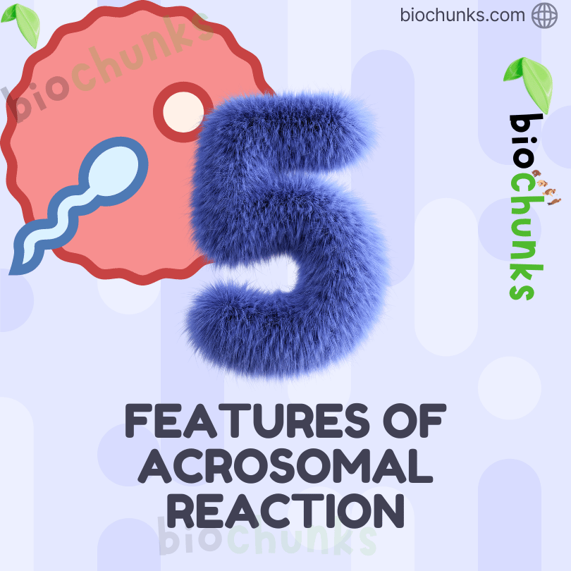
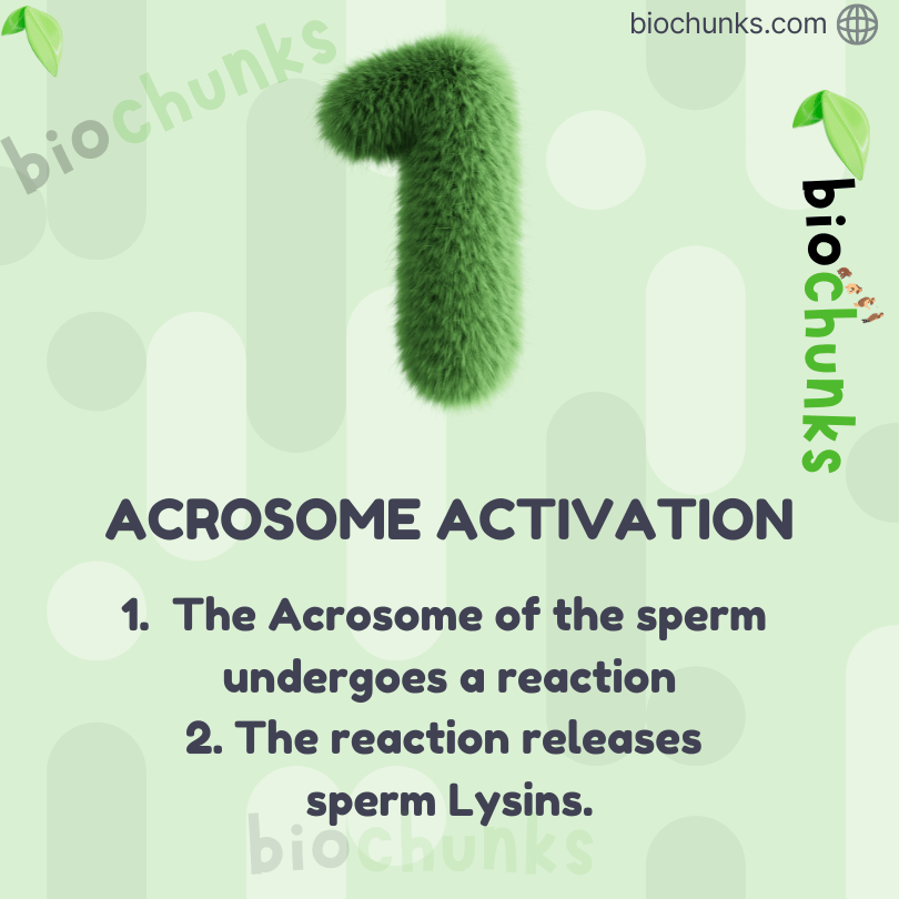
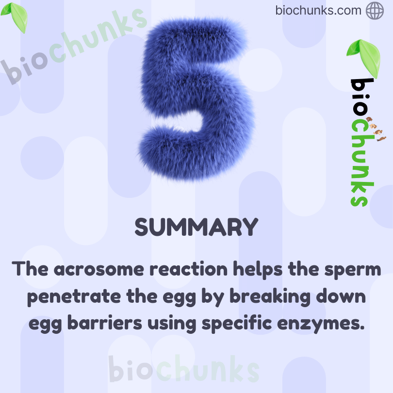
Acrosomal Reaction
- Definition
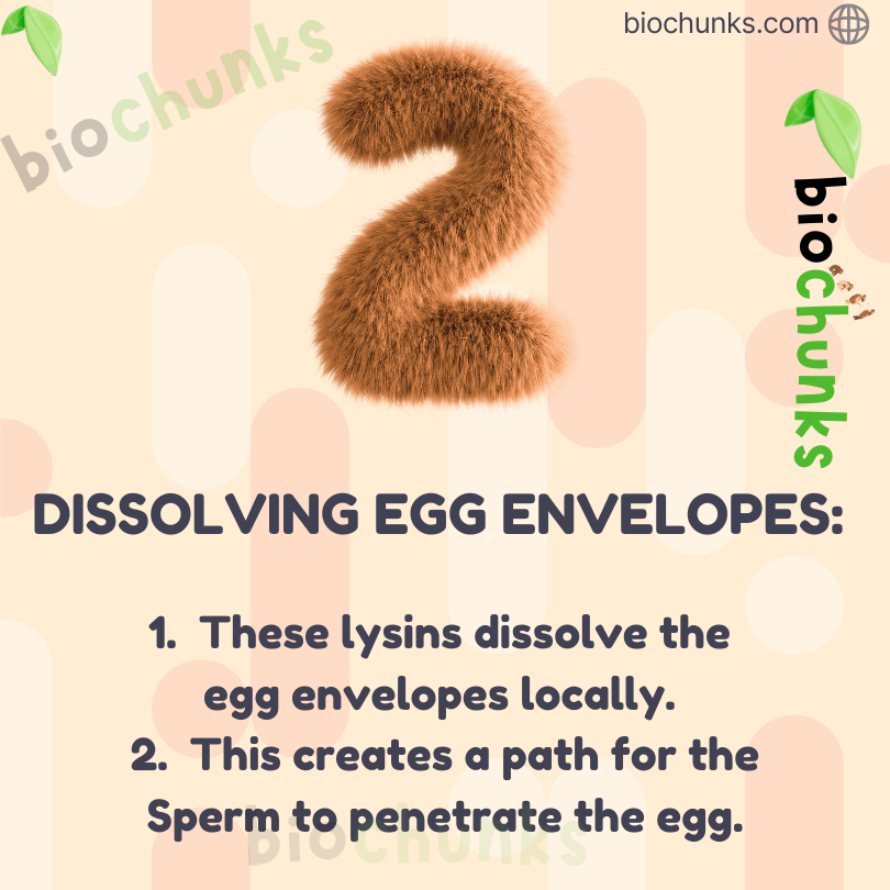
- Release of enzymes from the acrosome of the sperm to penetrate the ovum.
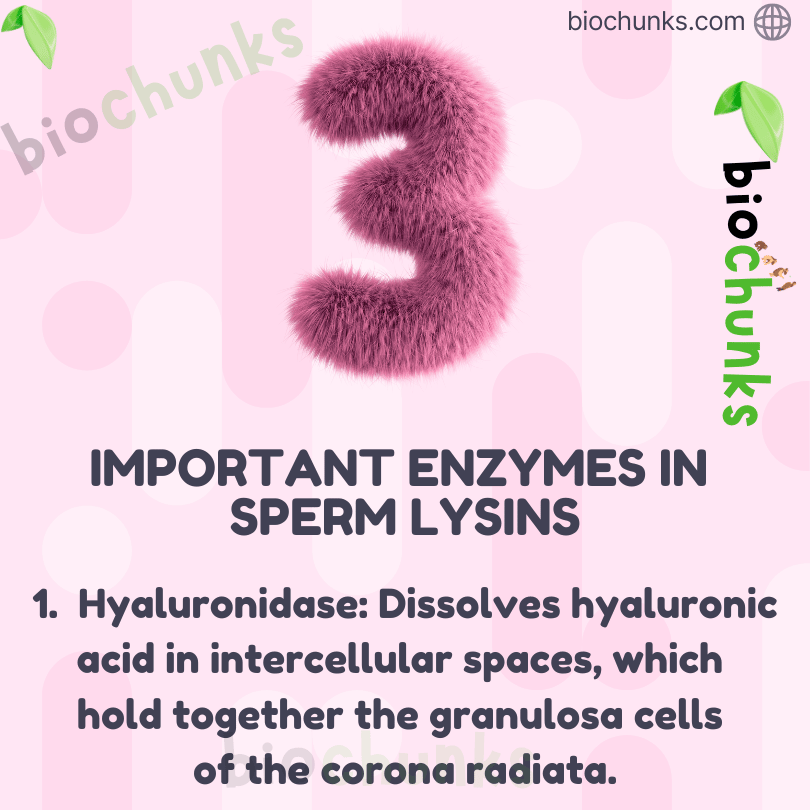
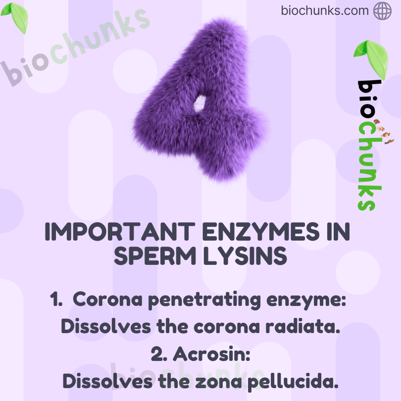
- Important Enzymes
- Hyaluronidase – dissolves ground substance of follicle cells.
- Corona penetrating enzymes – digest corona radiata.
- Acrosin – digests zona pellucida.
- Function
- Helps sperm enter the ovum.
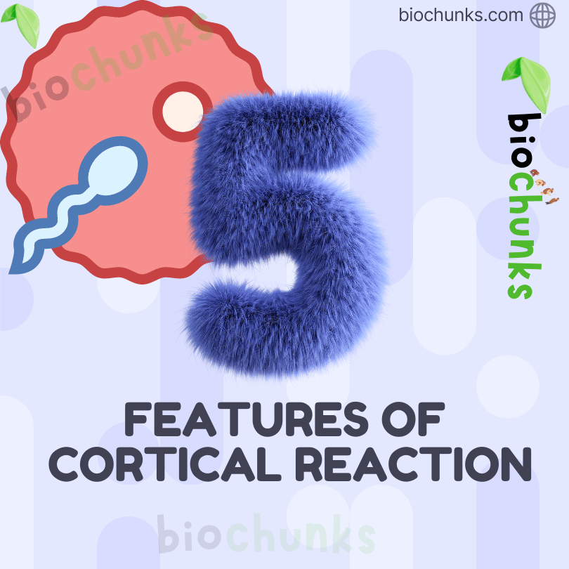
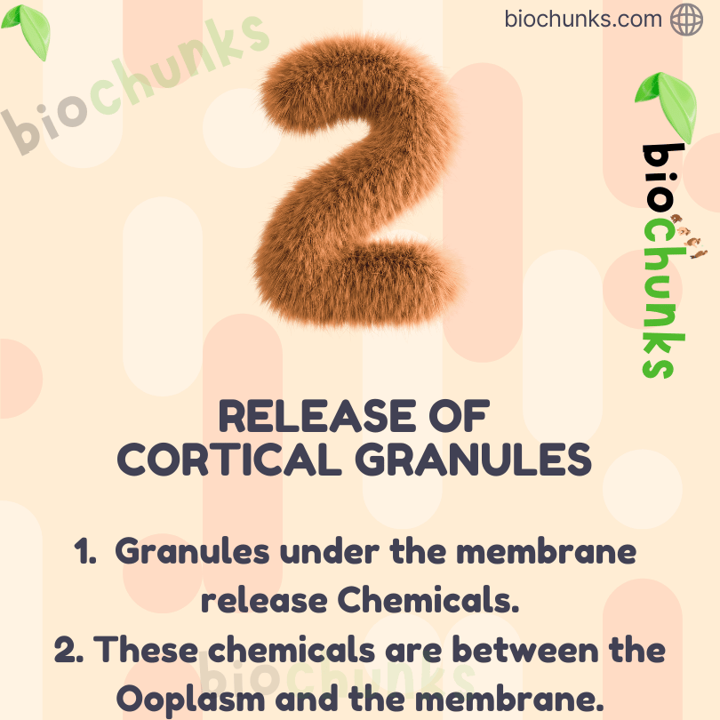
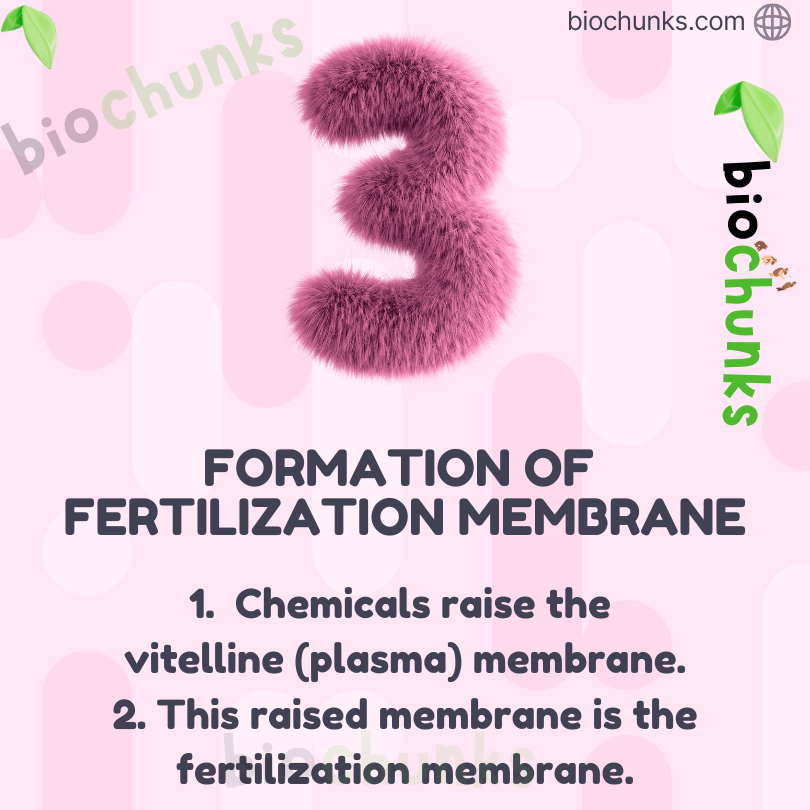
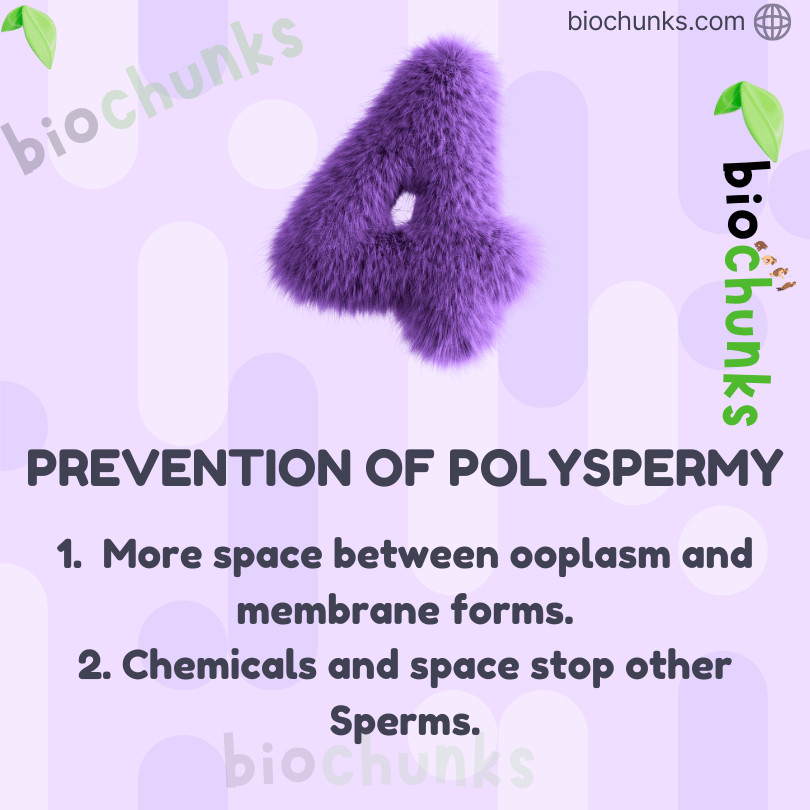
Role of Zona Pellucida and Cortical Reaction
- Zona Pellucida
- A protective glycoprotein layer around the ovum.
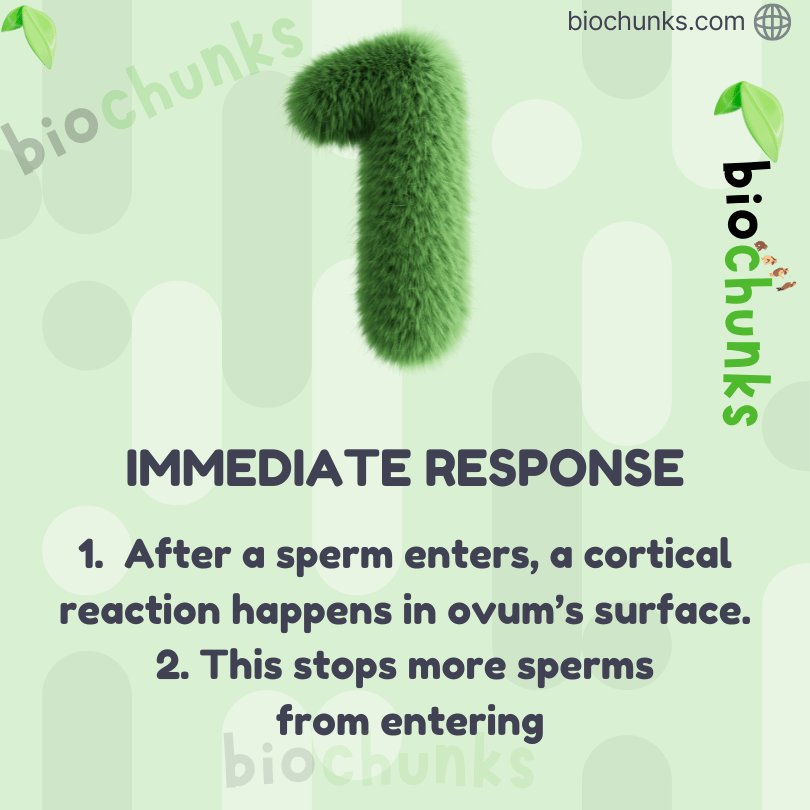
- Cortical Reaction
- Sperm contact with zona pellucida induces changes in ovum membrane.
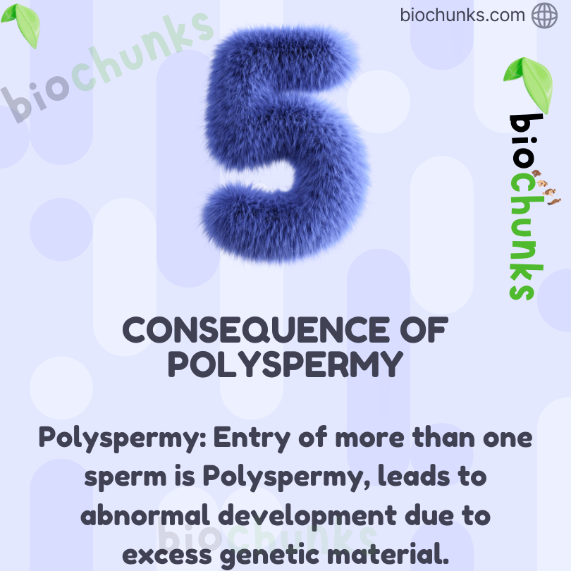
- Prevents entry of additional sperms.
- Ensures prevention of polyspermy.
Completion of Meiosis
- Event
- Entry of sperm induces completion of meiosis II in secondary oocyte.
- Result
- Formation of a haploid ovum.
- Formation of second polar body.
Zygote Formation
- Fusion
- Haploid sperm nucleus fuses with haploid ovum nucleus.
- Result
- Formation of diploid (2n) zygote.
Sex Determination
- Chromosome Patterns:
- Female: XX
- Male: XY
- Gametes Chromosomes:
- Female: All ova carry X chromosome.
- Male: Sperms carry either X or Y chromosome.
- Zygote Outcome:
- XX → Female child
- XY → Male child
- Key Point: Sex of the baby is determined by the father’s sperm.
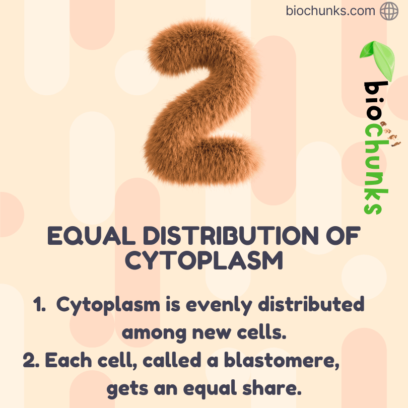
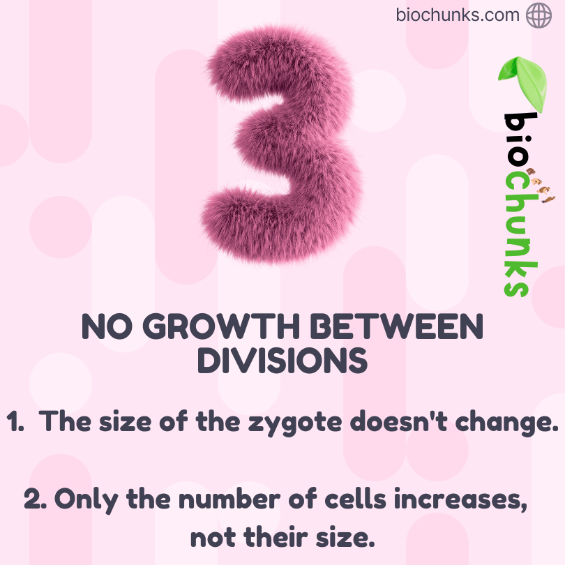
Cleavage and Blastocyst Formation

Cleavage
- Definition
- Series of rapid mitotic divisions of the zygote without increase in size.
- Location
- Occurs as the zygote moves through the oviduct toward the uterus.
- Stages
- 2-cell stage
- 4-cell stage
- 8-cell stage
- 16-cell stage (blastomeres)
- Type
- Holoblastic cleavage in humans.
Morula
- Definition
- Embryo with 8–16 blastomeres.
- Feature
- Solid ball of cells.
Blastocyst Formation
- Transformation
- Morula continues division and converts into blastocyst.
- Structure
- Trophoblast – outer cell layer.
- Inner Cell Mass – group of cells attached to trophoblast.
- Function
- Trophoblast helps in implantation.
- Inner cell mass forms the embryo.
Implantation
- Timing
- Occurs about 7 days after fertilisation.
- Process
- Blastocyst sheds zona pellucida (zona hatching).
- Trophoblast attaches to endometrium.
- Uterine cells proliferate rapidly.
- Blastocyst gets embedded in endometrium.
- Outcome
- Leads to pregnancy.
Significance of Implantation
- Ensures anchorage of embryo.
- Allows nutrient exchange with maternal tissues.
- Initiates embryonic development.


1.6 Pregnancy and Embryonic Development
Implantation and Placenta Formation
Implantation
- Implantation leads to pregnancy.
- After implantation, finger-like projections called chorionic villi appear on the trophoblast.
- These chorionic villi are surrounded by uterine tissue and maternal blood.
Placenta
- Placenta is formed by interdigitation of chorionic villi and uterine tissue.
- It forms a structural and functional connection between the embryo and the mother.
Functions of Placenta
- Supplies oxygen to the embryo.
- Supplies nutrients to the embryo.
- Removes carbon dioxide.
- Removes metabolic waste products.
Umbilical Cord
- Connects the embryo to the placenta.
- Helps in transport of nutrients, gases and wastes between embryo and mother.
Placenta as an Endocrine Tissue
- Hormones Secreted by Placenta:
- Human chorionic gonadotropin (hCG)
- Human placental lactogen (hPL)
- Estrogens and progestogens
Other Hormones During Pregnancy
- Relaxin: Relaxin is secreted by ovary during later stages of pregnancy.
- Increased Levels: of estrogens, progestogens, cortisol, prolactin and thyroxine increase in maternal blood.
Significance
- These hormones support fetal growth.
- Maintain pregnancy.
- Bring metabolic changes in the mother.
Embryonic Development
- Inner Cell Mass:
- Inner cell mass differentiates into three primary germ layers.
- Three Germ Layers
- Ectoderm – outer layer.
- Mesoderm – middle layer.
- Endoderm – inner layer.
- Importance
- All tissues and organs of the body develop from these three germ layers.
- Stem Cells
- Inner cell mass contains stem cells.
- Stem cells have the capacity to form all tissues and organs.

Stages of Embryonic Development
- 1 Month:
- Heart is formed.
- Heartbeat can be detected.
- 2 Months:
- Development of limbs and digits.
- 12 Weeks (End of First Trimester):
- Major organs are formed.
- Limbs are well developed.
- External genital organs are formed.
- 5 Months:
- First fetal movements are felt by the mother.
- Hair appears on the head.
- 24 Weeks (Second Trimester):
- Fine hair covers the body.
- Eyelids separate.
- Eyelashes form.
- 9 Months:
- Fetus is fully developed.
- Ready for birth.
Gestation Period
- Pregnancy duration is called gestation period.
- In humans, gestation period is about 9 months.
1.7 Parturition and Lactation
Parturition (Childbirth)
- Definition
- Parturition is the process of delivering the baby.
- Gestation Period
- Human pregnancy lasts about 9 months.
- Cause
- Strong, rhythmic contractions of the uterus.
- Nature
- A complex neuroendocrine process.
Trigger for Parturition
- Signals for Parturition
- Signals originate from the fully developed fetus and placenta.
- Foetal Ejection Reflex
- Initiates mild uterine contractions.
- Stimulates maternal pituitary gland.
- Oxytocin
- Released from maternal pituitary gland.
- Causes stronger uterine contractions.
- Positive Feedback
- Stronger contractions stimulate more oxytocin release.
- Continues until delivery of baby.
Delivery
- Baby is expelled through the birth canal.
- Placenta is expelled after birth (after-birth).
Lactation (Milk Production)
- Mammary Glands:
- Undergo differentiation during pregnancy.
- Start producing milk towards the end of pregnancy.
- Colostrum:
- First milk produced after childbirth.
- Yellowish in colour.
- Rich in antibodies.
- Importance of Colostrum
- Provides passive immunity to the newborn.
- Essential for development of immune system.
- Breastfeeding:
- Highly recommended for newborns.
- Provides nutrition and immunity.
Key Points (Quick Revision)
- Placenta acts as nutritive, respiratory, excretory and endocrine organ.
- hCG, hPL, estrogens and progestogens are pregnancy-specific hormones.
- Oxytocin plays a key role in inducing labour.
- Colostrum is antibody-rich and vital for newborn immunity.
Chapter Summary
Nature of Human Reproduction
- Humans are sexually reproducing and viviparous organisms, meaning fertilisation occurs inside the body and development takes place within the uterus, resulting in live birth.
Male Reproductive System: The male reproductive system consists of:
- A pair of testes (primary sex organs).
- Male sex accessory ducts.
- Accessory glands.
- External genitalia.
- Structure and Function of Testes
- Each testis contains about 250 compartments called testicular lobules.
- Each lobule has one to three highly coiled seminiferous tubules.
- Cells of Seminiferous Tubules
- Spermatogonia: Undergo meiotic divisions to form sperms.
- Sertoli cells: Provide nourishment and support to developing germ cells.
- Interstitial Cells
- Leydig cells are present outside seminiferous tubules.
- They synthesize and secrete male sex hormones called androgens.
- Male External Genitalia
- The external genital organ in males is the penis, which plays a role in insemination.
Female Reproductive System: The female reproductive system includes:
- A pair of ovaries.
- A pair of oviducts (fallopian tubes).
- A uterus.
- A vagina.
- External genitalia.
- A pair of mammary glands.
- Functions of Ovaries
- Produce female gametes (ova).
- Secrete steroid hormones called ovarian hormones.
- Ovarian follicles at different developmental stages are present in the ovarian stroma.
- Female Accessory Ducts
- Include the oviducts, uterus and vagina.
- Structure of Uterus: The uterine wall has three layers:
- Perimetrium – outer protective layer.
- Myometrium – middle muscular layer.
- Endometrium – inner glandular layer that undergoes cyclic changes.
- Female External Genitalia
- Include mons pubis, labia majora, labia minora, hymen and clitoris.
- Mammary Glands
- Mammary glands are secondary sexual characteristics in females.
- They differentiate during pregnancy and produce milk after childbirth.
Gametogenesis
- Spermatogenesis is the process of formation of sperms in males.
- Oogenesis is the process of formation of mature female gametes (ova).
Structure of Human Sperm
- A normal human sperm consists of a head, neck, middle piece and tail.
Menstrual Cycle
- The reproductive cycle in female primates is called the menstrual cycle.
- It begins at puberty and continues until menopause.
- Usually, one ovum is released per menstrual cycle during ovulation.
- Cyclical changes in ovary and uterus are regulated by pituitary and ovarian hormones.
Fertilisation and Development
- After coitus, sperms reach the ampullary–isthmic junction of the oviduct.
- Fertilisation occurs at this site, resulting in the formation of a diploid zygote.
- The sex of the embryo is determined by whether the sperm carries an X or Y chromosome.
Cleavage and Implantation
- The zygote undergoes mitotic divisions to form a blastocyst.
- The blastocyst implants in the uterine wall, leading to pregnancy.
Pregnancy and Birth
- Pregnancy lasts about nine months (gestation period).
- The fully developed fetus is ready for delivery at the end of gestation.
Parturition
- Childbirth or parturition is induced by a complex neuroendocrine mechanism.
- Hormones like cortisol, estrogens and oxytocin play a key role.
Lactation
- After childbirth, mammary glands secrete milk.
- The newborn is nourished by breast milk during the initial months of growth.
- This process is called lactation.


